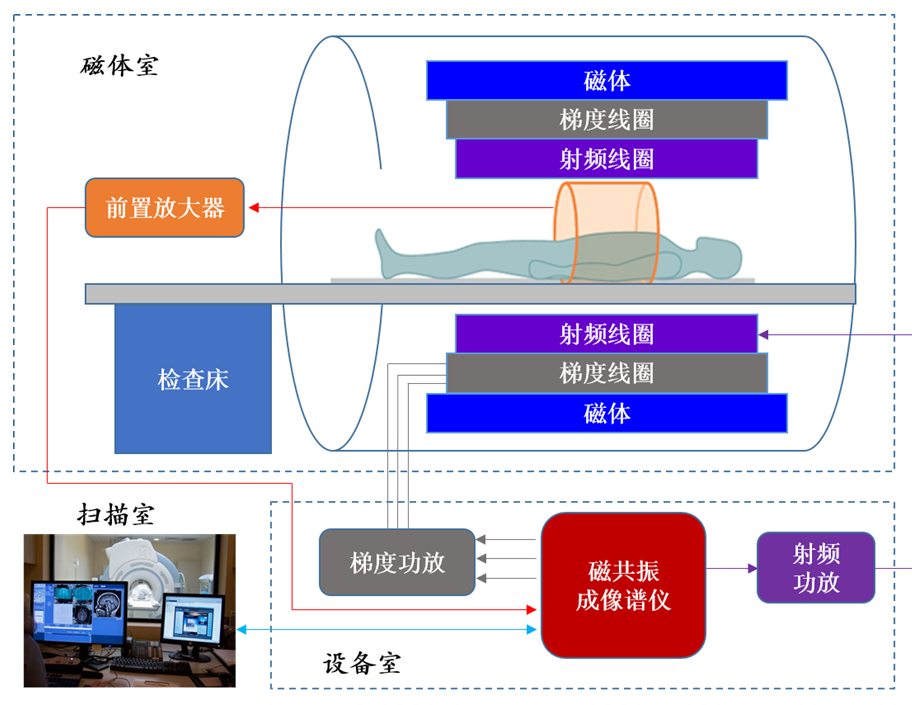
Medical magnetic resonance imaging (MRI) technology is an important application field of nuclear magnetic resonance (NMR). MRI technology can image any tissue of the body, recording its relaxation, proton density, flow, chemical shifts, diffusion, perfusion, blood oxygenation status, temperature and so on. It has very important value in the inspection of brain and spinal cord. Because magnetic resonance imaging can observe the pathological changes in three dimensional space, it has become an important preoperative location method, which is conducive to the diagnosis, treatment and curative effect of the tumor. MRI also has the advantages of high soft tissue resolution, arbitrary cross-section imaging and non-ionizing radiation. It’s one of the most advanced diagnostic techniques in the field of medical imaging. MRI system mainly includes the following parts: spectrometer system, pulse sequence design and so on.

1. Spectrometer system
MRI spectrometer is the physical platform for the operation and implementation of the imaging sequences: Firstly Spectrometer operates the pulse sequences downloaded from the user computer, generating the RF and gradient signal in accordance with the specified and precise timing sequence (driving corresponding coils to generate RF and gradient field after amplified), then demodulates and filters the MR signal detected from the receiving coil, finally will upload the data back to the user computer to rebuild the image. Spectrometer is the core of the whole MRI system, its performance plays a decisive role in imaging function and quality of MRI system. In recent years, new technologies of MR spectrum instrument emerge in an endless stream, from the beginning double channel, the RF system has developed to the present 8 to 32 channels even 64 channels, and the dual and four source emission technology also emerge. These technologies combined with parallel imaging technology, greatly shorten the scanning time and improve the signal to noise ratio and image uniformity.
(1) Optical fiber spectrometer system
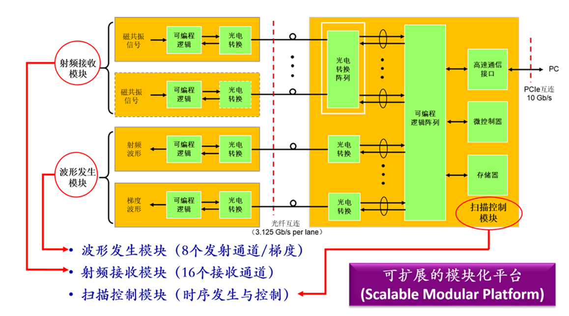
Our laboratory has been performing research on the key technologies of the digital optical fiber spectrometer for high field (1.5T and 3T) magnetic resonance imaging systems. Through advanced digital technology and new-type rapid prototyping (and 3D printing technology) to solve the core problem of optical fiber in RF system. Through the theory analysis and algorithm design, combining the related experimental verification to develop the key technologies such as multi-source emission (2~8 emission channel), fast imaging, high-speed data processing and transmission; combining the clinical application problems on real-time imaging, parallel imaging and functional imaging, perform study on the hardware and software of MRI spectrometer, ultrafast echo technique and whole body imaging technology, to provide strong guidance and support for the future development of ultra-high field (7T and 11T) high performance MRI spectrometer system, promoting MRI technology and industrialization in our country.

This platform can achieve at least eight parallel transmission channel and 32 parallel receive channels, flexible, precise gradient control, real-time image reconstruction and data transmission function. Ensuring the spectrometer has the advantages of scalability, high performance and high reliability, line number for system interconnection, electromagnetic interference, the complexity and cost of the hardware circuit design are all reduced.
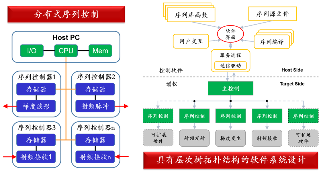
(2) Spectrometer software and sequence control
Spectrometer software is mainly divided into five parts: user interface software, pulse sequence, sequence function library, and start program for sequence running. The user interface software, pulse sequence and function library are written with Visual C++ 2010 and run on the windows platform of the host computer, calling the PCIe driver to achieve the data exchange between data transmission service program in user interface software and data communication modules, and the spectrometer performs the corresponding operation. Before the sequence operating, user interface software will call sequence program written by user to generate binary file (bin file) sending to the spectrometer.

2. Pulse sequence
MRI pulse sequence is actually the arrangement of radio frequency pulse and gradient field changes in the time sequence, so each pulse sequence will involve the repetition time, echo time, effective echo time, echo train length, echo spacing, reversal time, excitation frequency, acquisition time and other key concepts.
